This special page shows all files uploaded to the English Wikipedia. (See also the list for Wikimedia Commons.) By default, the last uploaded files are shown at top of the list, but clicking on a column header will change the sorting.
Deleted files are not shown here but are included in the upload log.
| Date | Name | Thumbnail | Size | Description |
|---|---|---|---|---|
| 07:01, 23 October 2009 | Esophageal varices being banded, showing white ball sign and wale sign.JPG ( file) |
 |
87 KB | {{Information |Description = Endoscopy image of bleeding esophageal varices being banded. Clearly seen are the longitudinal wale signs on the banded varix. Note the colour change in the banded varix to an off-white shade, indicating that it is d |
| 08:57, 8 October 2007 | Eosinophilic esophagitis endo.jpg ( file) |
 |
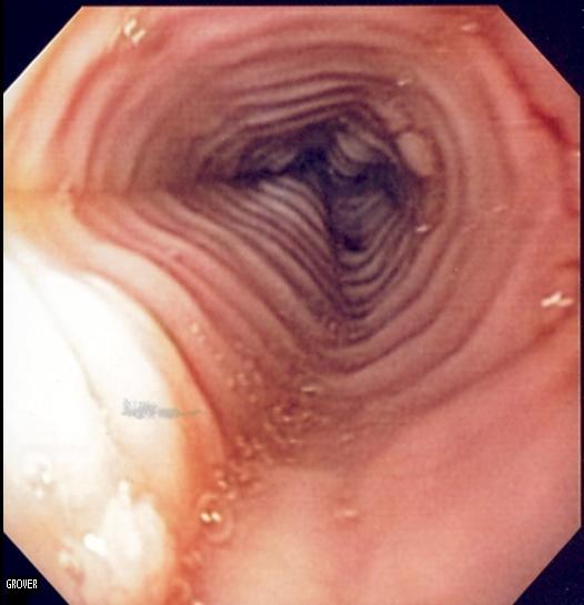
28 KB | == Summary == Endoscopic image of patient with eosinophilic esophagitis showing multiple rings and linear furrows. Signed release into public domain from patient. -- Samir 08:37, 8 October 2007 (UTC) == Licensing == {{self|GFDL-no-disc |
| 05:24, 15 May 2007 | Postpolypectomy ulcer.jpg ( file) |
 |
245 KB | Colonoscopic image of ulcer seen bleeding after polypectomy, closed with an endoclip. Released into public domain on permission of patient. -- ~~~~ Category:Endoscopic images |
| 05:22, 15 May 2007 | Resolution clip.jpg ( file) |
 |
843 KB | Photograph of a single-use rotatable endoclip (Resolution Clip, Boston Scientific Inc.) -- ~~~~ |
| 06:52, 19 January 2007 | Fluoro biliary SEMS.jpg ( file) |
 |
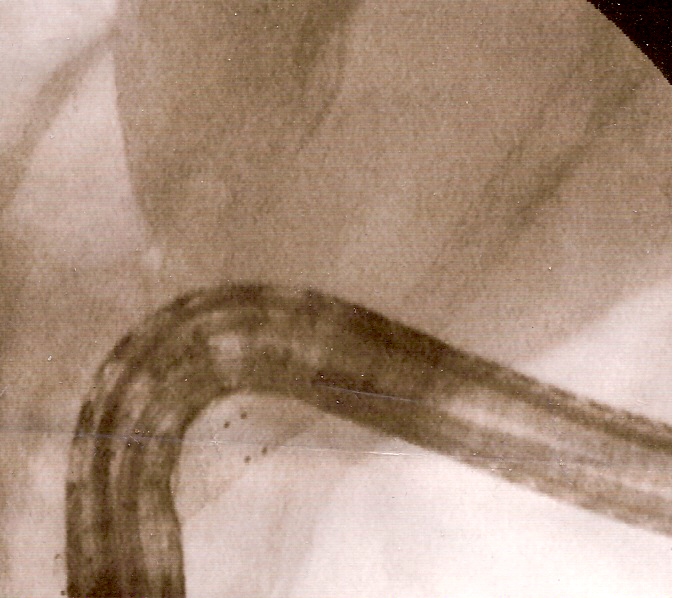
147 KB | Fluoroscopic image of two metal biliary stents in the common bile duct. The large black tube across the image is the duodenoscope. Released into public domain on written permission of patient -- ~~~~ [[Category:En |
| 06:26, 19 January 2007 | ERCP DACP stones.jpg ( file) |
 |
97 KB | Fluoroscopic image of multiple common bile duct stones seen at the time of ERCP and duodenoscope assisted cholangiopancreatography (DACP]]. The stone was impacted in the d |
| 21:11, 8 October 2006 | Sarin classification.jpg ( file) |
 |
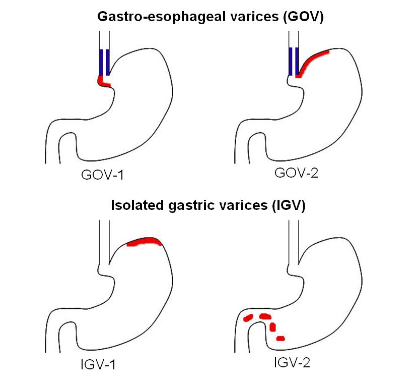
41 KB | Schematic of Sarin classification of gastric varices, adapted from {{cite journal | author = Sarin S | title = Long-term follow-up of gastric variceal sclerotherapy: an eleven-year experience. | journal = Gastrointest Endosc | volume = 46 | issue = 1 |
| 16:29, 12 September 2006 | Labelled coeliac path.jpg ( file) |
 |
154 KB | Labelled coeliac pathology showing villi, crypts, lymphocytes -- ~~~~ |
| 05:00, 9 September 2006 | Eosinophilic esophagiits ba swallow.jpg ( file) |
 |
15 KB | Barium swallow of multi ring esophagus of eosinophilic esophagitis, taken from Commons immage Image:Eosinophilic_esophagitis-barium_swallow.jpg and modified to remove achalasia barium swallow and to remove mark on X-ray film -- ~~~~ |
| 09:46, 8 September 2006 | Infliximab structure.jpg ( file) |
 |
19 KB | Reverted to earlier revision |
| 11:15, 31 August 2006 | Cytokines in IBD.jpg ( file) |
 |
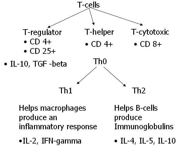
36 KB | Schematic demonstrating the cytokines involved in inflammatory bowel disease pathogenesis -- ~~~~ |
| 07:20, 15 August 2006 | Impacted ampulla.jpg ( file) |
 |
51 KB | Endoscopic still of common bile duct stone impacted at ampulla of Vater. Released into public domain on permission of patient. -- ~~~~ Category:Endoscopic images |
| 11:26, 13 August 2006 | Eosinophilic esophagiits path.jpg ( file) |
 |
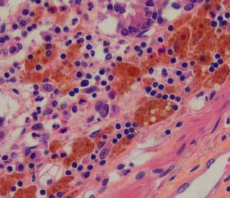
60 KB | H&E stain of esophagus biopsy showing eosinophilic esophagitis, manifested by an infiltration of eosinophils in the lamina propria. -- ~~~~ |
| 05:27, 28 July 2006 | Melanosis path.jpg ( file) |
 |
36 KB | Biopsy of colon showing melanosis coli, which appears as brown pigmentation in the lamina propria. Released with permission by patient into public domain -- ~~~~ |
| 06:56, 7 July 2006 | Edith Sampson.jpg ( file) |
 |
29 KB | {{c-uploaded}} For main page from Commons -- ~~~~ |
| 11:00, 7 June 2006 | Imuran 2.JPG ( file) |
 |
705 KB | Better picture of azathioprine tablets, taken by myself -- ~~~~ |
| 06:19, 6 June 2006 | Coin in esophagus lat 2.jpg ( file) |
 |
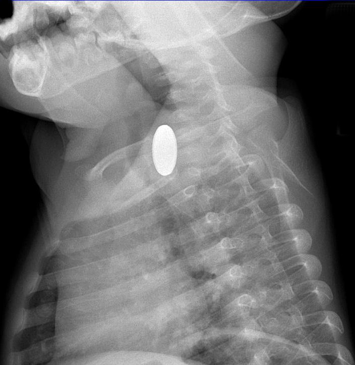
70 KB | Lateral view Chest X-Ray of Canadian dollar coin in esophagus of child. Released per permission of mother. -- ~~~~ |
| 22:53, 5 June 2006 | Brain met.jpg ( file) |
 |
50 KB | CT scan of breast adenoca metastasized to L parietal area peri-ventricular area. Released into public domain on permission of patient. -- ~~~~ |
| 20:49, 3 June 2006 | CBC with Hct.jpg ( file) |
 |
10 KB | Physician shorthand for CBC -- ~~~~ |
| 14:08, 3 June 2006 | Cortifoam.jpg ( file) |
 |
119 KB | Image of Cortifoam enema -- ~~~~ |
| 12:54, 3 June 2006 | NOD2 CARD15.jpg ( file) |
 |
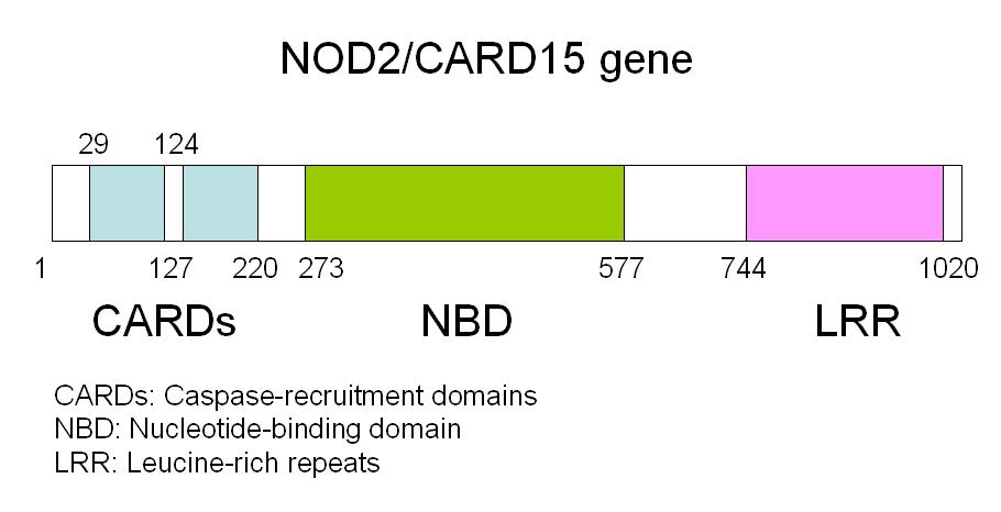
38 KB | Schematic of NOD2 CARD15 gene -- ~~~~ |
| 11:15, 3 June 2006 | CBC and lytes schematic.jpg ( file) |
 |
8 KB | Schematic for shorthand for complete blood count and electrolytes measurements in blood used in North America -- ~~~~ |
| 09:06, 3 June 2006 | Patterns of CD.jpg ( file) |
 |
45 KB | Schematic of patterns of disease in CD. Based on diagram of human intestine in GFDL. -- ~~~~ |
| 09:00, 3 June 2006 | Colonic CD.png ( file) |
 |
59 KB | Based on diagram of human intestine, shaded colon in patchy distribution to indicate colonic CD -- ~~~~ |
| 09:00, 3 June 2006 | Ileocolic CD.png ( file) |
 |
60 KB | Based on diagram of human intestine, shaded ileum and patchy colon to demonstrate ileocolic CD -- ~~~~ |
| 08:59, 3 June 2006 | Ileal CD.png ( file) |
 |
53 KB | Based on diagram of human intestine, shaded ileum to demonstrate CD -- ~~~~ |
| 07:43, 2 June 2006 | SEMS fluoro 2.jpg ( file) |
 |
21 KB | Fluoroscopic image of patent self-expanding metallic stent in esophagus. Released into public domain on permission of patient. -- ~~~~ |
| 05:53, 17 May 2006 | Glycogenic acanthosis.jpg ( file) |
 |
25 KB | Endoscopic image of glycogenic acanthosis, placed in public domain with permission of patient -- ~~~~ |
| 04:53, 17 May 2006 | Gastric varices.jpg ( file) |
 |
48 KB | Isolated gastric varices seen on retroflexion of the gastroscope. The picture was placed into the public domain on the permission of the patient. -- ~~~~ |
| 00:45, 5 May 2006 | Gastric ulcer.jpg ( file) |
 |
42 KB | Endoscopic picture of gastric ulcer with visible vessel. Reproduced with permission of patient. -- ~~~~ |
| 02:26, 3 May 2006 | Moe Contribs.jpg ( file) |
 |
44 KB | Moe Epsilon's last contribs -- ~~~~ |
| 16:36, 2 May 2006 | Diverticulosis 1.jpg ( file) |
 |
39 KB | Endoscopic image of diverticulosis, taken by myself, and released with permission of patient into public domain -- ~~~~ |
| 04:35, 20 April 2006 | Canadian maple leaf 2.jpg ( file) |
 |
73 KB | Better image of leaf of Canadian Maple photoshopped to illustrate detail -- ~~~~ |
| 04:30, 20 April 2006 | Canadian maple leaf.jpg ( file) |
 |
305 KB | Image of leaf of Canada maple, photoshopped to illustrate detail -- ~~~~ |