| Hamate bone | |
|---|---|
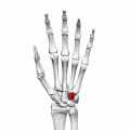
_01_palmar_view.png) Left hand anterior view (palmar view). Hamate bone shown in red. | |
 The left hamate bone | |
| Details | |
| Pronunciation | /ˈheɪmət/ |
| Articulations | Articulates with five bones: the lunate proximally the fourth and fifth metacarpals distally the triangular medially the capitate laterally |
| Identifiers | |
| Latin | os hamatum |
| MeSH | D051225 |
| TA98 | A02.4.08.012 |
| TA2 | 1259 |
| FMA | 23730 |
| Anatomical terms of bone | |
The hamate bone (from Latin hamatus, "hooked"), or unciform bone (from Latin uncus, "hook"), Latin os hamatum and occasionally abbreviated as just hamatum, [1] [2] [3] is a bone in the human wrist readily distinguishable by its wedge shape and a hook-like process ("hamulus") projecting from its palmar surface.
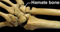
The hamate is an irregularly shaped carpal bone found within the hand. The hamate is found within the distal row of carpal bones, and abuts the metacarpals of the little finger and ring finger. [4]: 708–709
Adjacent to the hamate on the ulnar side, and slightly above it, is the pisiform bone. Adjacent on the radial side is the capitate, and proximal is the lunate bone. [4]: 708–709
The hamate bone has six surfaces:
- The superior, the apex of the wedge, is narrow, convex, smooth, and articulates with the lunate.
- The inferior articulates with the fourth and fifth metacarpal bones, by concave facets which are separated by a ridge.
- The dorsal is triangular and rough for ligamentous attachment.
- The palmar presents, at its lower and ulnar side, a curved, hook-like process, the hamulus, directed forward and laterally.
- The medial articulates with the triangular bone by an oblong facet, cut obliquely from above, downward and medialward.
- The lateral articulates with the capitate by its upper and posterior part, the remaining portion being rough, for the attachment of ligaments.
_-_animation02.gif)
The hook of hamate ( Latin: hamulus) is found at the proximal, ulnar side of the hamate bone. The hook is a curved, hook-like process that projects 1–2 mm distally and radially. [5] The ulnar nerve hooks around the hook of hamate as it crosses towards the medial side of hand.
The hook forms the ulnar border of the carpal tunnel, and the radial border for Guyon's canal. Numerous structures attach to it, including ligaments from the pisiform, the transverse carpal ligament, and the tendon of flexor carpi ulnaris. [5]
Its medial surface to the flexor digiti minimi brevis and opponens digiti minimi; its lateral side is grooved for the passage of the flexor tendons into the palm of the hand.
The ossification of the hamate starts between 1 and 12 months. [6] The hamate does not fully ossify until about the 15th year of life. [5]
The bone is also found in many other mammals, and is homologous with the "fourth distal carpal" of reptiles and amphibians.
|
| This section needs expansion. You can help by
adding to it. (September 2022) |
The carpal bones function as a unit to provide a bony superstructure for the hand. [4]: 708
The hamate bone is the bone most commonly fractured when a golfer hits the ground hard with a golf club on the downswing or a hockey player hits the ice with a slap shot. The fracture is usually a hairline fracture, commonly missed on normal X-rays. Symptoms are pain aggravated by gripping, tenderness over the hamate and symptoms of irritation of the ulnar nerve. This is characterized by numbness and weakness of the fifth digit with partial involvement of the fourth digit as well, the "ulnar 1½ fingers".
The hook of hamate is particularly prone to fracture-related complications such as non-union due to its tenuous blood supply. [5]
It is also a common injury in baseball players. Several professional baseball players have had the bone removed during the course of their careers. [7] [8] [9] [10] [11] [12] This condition has been called "Wilson's Wrist". [13]
The calcification of the hamate bone is seen on X-rays during puberty and is sometimes used in orthodontics to determine if an adolescent patient is suitable for orthognathic intervention (i.e. before or at their growth spurt).[ citation needed]
The etymology derives from the Latin hamatus "hooked," from hamus which means "hook".
-
 Position of hamate bone (shown in red). Left hand. Animation.
 -
 Hamate bone of the left hand. The hook-like process is called hamulus.
 -
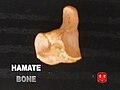 Hamate bone.
 -
 Right hand anterior view (palmar view). Thumb on top.
 -
 Right hand posterior view (dorsal view). Thumb on bottom.
 -
 Bones of the left hand. Palmar surface. Hamate shown in yellow.
 -
 Bones of the left hand. Dorsal surface. Hamate shown in yellow.
 -
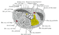 Transverse section across the wrist and digits. Hamate shown in yellow.
 -
 Cross section of wrist (thumb on left). Hamate shown in red.
 -
 Right wrist joint. Deep dissection. Anterior (palmar) view.
_-_animation01.gif)
_-_animation04.gif)


![]() This article incorporates text in the
public domain from
page 227 of the 20th edition of
Gray's Anatomy (1918)
This article incorporates text in the
public domain from
page 227 of the 20th edition of
Gray's Anatomy (1918)
- ^ Gdoura, F.; Trigui, M.; Ellouze, Z.; Hamed, Y. B.; Ayadi, K.; Keskes, H. (October 2010). "Hamatum osteoblastoma". Orthopaedics & Traumatology, Surgery & Research: OTSR. 96 (6): 712–716. doi: 10.1016/j.otsr.2010.02.014. ISSN 1877-0568. PMID 20692218.
- ^ Mei, Guo-Hua; Wang, Hai-Ming; Fan, Cun-Yi; Zhang, Chang-Qing; Zeng, Bing-Fang (October 2014). "Possibility of the hamatum carpometacarpal joint as a new joint donor site for interphalangeal joint restoration". European Journal of Orthopaedic Surgery & Traumatology: Orthopedie Traumatologie. 24 (7): 1175–1180. doi: 10.1007/s00590-013-1300-4. ISSN 1633-8065. PMID 23982116. S2CID 25697262.
- ^ Alp, Nazmi Bülent; Kaleli, Tufan; Kalay, Onur Can; Karpat, Fatih; Akdag, Gokhan; Macunluoglu, Aslı Ceren; Oral, Gamze Saygı (2020). "The Effect of Hamatum Curvature Angle on Carpal Tunnel Volumetry: A Mathematical Simulation Model". Computational and Mathematical Methods in Medicine. 2020: 7582181. doi: 10.1155/2020/7582181. ISSN 1748-6718. PMC 7312712. PMID 32617118.
- ^ a b c Drake, Richard L.; Vogl, Wayne; Tibbitts, Adam W.M. Mitchell; illustrations by Richard; Richardson, Paul (2005). Gray's anatomy for students. Philadelphia: Elsevier/Churchill Livingstone. ISBN 978-0-8089-2306-0.
- ^ a b c d Eathorne, SW (March 2005). "The wrist: clinical anatomy and physical examination—an update". Primary Care. 32 (1): 17–33. doi: 10.1016/j.pop.2004.11.009. PMID 15831311.
- ^ Balachandran, Ajay; Kartha, Moumitha; Krishna, Anooj; Thomas, Jerry; K, Prathilash; TN, Prem; GK, Libu; B, Krishnan; John, Liza (2014). "A Study of Ossification of Capitate, Hamate, Triquetral & Lunate in Forensic Age Estimation". Indian Journal of Forensic Medicine & Toxicology. 8 (2): 218–224. doi: 10.5958/0973-9130.2014.00720.8. ISSN 0973-9130. Retrieved 18 August 2014.
- ^ Snow, Chris (June 1, 2006). "Peña to have surgery". The Boston Globe. Retrieved September 2, 2011.
- ^ Manuel, John (March 31, 2004). "Wrist Troubles Drain Prospects' Power". Baseball America. Retrieved September 2, 2011.
- ^ Benjamin, Amalie (July 27, 2007). "He's gaining in arms race". The Boston Globe. Retrieved September 2, 2011.
- ^ "Dickerson has hand, wrist surgery". ESPN. Associated Press. May 3, 2010. Retrieved September 2, 2011.
- ^ Carobine, Kieran (March 8, 2011). "Domonic Brown's Surgery A Success". Phillies Nation. Retrieved September 2, 2011.
- ^ "Angels' Mike Trout: Undergoes hamate surgery". CBS Sports. July 5, 2023. Retrieved August 4, 2023.
- ^ WILSON JN. Profiles of the carpal canal. J Bone Joint Surg Am. 1954 Jan;36-A(1):127–132