
Size of this preview:
600 × 600 pixels. Other resolutions:
240 × 240 pixels |
480 × 480 pixels |
768 × 768 pixels |
1,024 × 1,024 pixels |
1,411 × 1,411 pixels.
Original file (1,411 × 1,411 pixels, file size: 248 KB, MIME type: image/jpeg)
| This is a file from the
Wikimedia Commons. Information from its
description page there is shown below. Commons is a freely licensed media file repository. You can help. |
Summary
| Description |
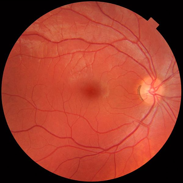
English:
Fundus photograph of the right eye, showing a
fundus with no sign of disease or pathology. It is seen from front so that left in each image is to the person's right. The gaze is into the camera, so the
macula is in the center of the image, and the
optic disk is located towards the nose (right in image). The optic disk has some pigmentation at the perimeter of the lateral side, which is considered non-pathological.
Veins are darker and slightly wider than corresponding arteries. Major nerve pathways are seen as white striped patterns radiating from the optic disk. In addition, there are also lighter areas close to larger vessels seen mainly at upper left in the image (person's upper right), which is regarded as a normal finding in younger people. Photo is taken at Gävle Hospital in Sweden in 2012 on a healthy 25-year old male volunteer. |
| Date | |
| Source | Own work |
| Author |
When using this image in external works, it may be cited as:
or
|
| Other versions |
 |
Licensing
I, the copyright holder of this work, hereby publish it under the following license:
| This file is made available under the Creative Commons CC0 1.0 Universal Public Domain Dedication. | |
| The person who associated a work with this deed has dedicated the work to the
public domain by waiving all of their rights to the work worldwide under copyright law, including all related and neighboring rights, to the extent allowed by law. You can copy, modify, distribute and perform the work, even for commercial purposes, all without asking permission.
|
File history
Click on a date/time to view the file as it appeared at that time.
| Date/Time | Thumbnail | Dimensions | User | Comment | |
|---|---|---|---|---|---|
| current | 10:49, 21 March 2012 |
| 1,411 × 1,411 (248 KB) | Mikael Häggström |
File usage
The following pages on the English Wikipedia use this file (pages on other projects are not listed):
Global file usage
The following other wikis use this file:
- Usage on ar.wikipedia.org
- Usage on bs.wikipedia.org
- Usage on ca.wikipedia.org
- Usage on de.wikipedia.org
- Usage on en.wikiversity.org
- Usage on et.wikipedia.org
- Usage on fr.wikipedia.org
- Usage on fr.wikiversity.org
- Usage on gl.wikipedia.org
- Usage on he.wikipedia.org
- Usage on hu.wikipedia.org
- Usage on it.wikipedia.org
- Usage on ml.wikipedia.org
- Usage on pl.wikipedia.org
- Usage on simple.wikipedia.org
- Usage on th.wikipedia.org
- Usage on tr.wikipedia.org
- Usage on uk.wikipedia.org
- Usage on zh-yue.wikipedia.org
- Usage on zh.wikipedia.org