Size of this preview:
728 × 600 pixels. Other resolutions:
291 × 240 pixels |
583 × 480 pixels |
930 × 766 pixels.
Original file (930 × 766 pixels, file size: 904 KB, MIME type: image/png)
| This is a file from the
Wikimedia Commons. Information from its
description page there is shown below. Commons is a freely licensed media file repository. You can help. |
Summary
| Description |
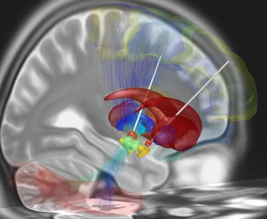
English: Depicted is a reconstruction of bihemispheric DBS electrodes that have been surgically placed into the most common target structure for treatment of Parkinson's Disease, the subthalamic nucleus (orange). Other subcortical structures include the red nucleus (green), the substantia nigra (yellow), the internal (cyan) and external (blue) pallidum and the striatum (red). A stimulation volume is modeled by applying 2V (at 1000Ω impedance) to the second-uppermost contact of the left electrode. Structural fibertracts traversing through this volume are visualized and cortical regions that they connect with the stimulation volume are selected from an automatic anatomical labeling atlas and visualized.
Picture created using LEAD DBS software (www.lead-dbs.org) |
| Date | |
| Source | Own work |
| Author | Andreashorn |
Licensing
I, the copyright holder of this work, hereby publish it under the following license:
This file is licensed under the
Creative Commons
Attribution-Share Alike 4.0 International license.
- You are free:
- to share – to copy, distribute and transmit the work
- to remix – to adapt the work
- Under the following conditions:
- attribution – You must give appropriate credit, provide a link to the license, and indicate if changes were made. You may do so in any reasonable manner, but not in any way that suggests the licensor endorses you or your use.
- share alike – If you remix, transform, or build upon the material, you must distribute your contributions under the same or compatible license as the original.
File history
Click on a date/time to view the file as it appeared at that time.
| Date/Time | Thumbnail | Dimensions | User | Comment | |
|---|---|---|---|---|---|
| current | 20:06, 16 May 2015 |
| 930 × 766 (904 KB) | Defaultmode42 | User created page with UploadWizard |
File usage
The following pages on the English Wikipedia use this file (pages on other projects are not listed):
Global file usage
The following other wikis use this file:
- Usage on de.wikipedia.org
- Usage on en.wikiversity.org
- Usage on fr.wikipedia.org
- Usage on sr.wikipedia.org