
Size of this preview:
800 × 549 pixels. Other resolutions:
320 × 220 pixels |
640 × 439 pixels |
1,024 × 703 pixels |
1,400 × 961 pixels.
Original file (1,400 × 961 pixels, file size: 959 KB, MIME type: image/png)
| This is a file from the
Wikimedia Commons. Information from its
description page there is shown below. Commons is a freely licensed media file repository. You can help. |
| Description |
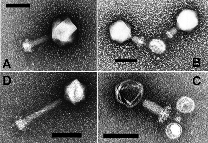
Electron Micrograph of Negative-Stained Prochlorococcus Myoviruses P-SSM2 and P-SSM4. Myovirus P-SSM2 with (A) non-contracted tail and (B) contracted tail, and myovirus P-SSM4 with (C) contracted tail and (D) non-contracted tail. Note the T4-like capsid, baseplate, and tail structure in both myoviruses. Scale bars indicate 100 nm. |
||
| Date | Published: April 19, 2005 | ||
| Source | Sullivan MB, Coleman ML, Weigele P, Rohwer F, Chisholm SW. Three Prochlorococcus Cyanophage Genomes: Signature Features and Ecological Interpretations. PLoS Biology Vol. 3/5/2005, e144 doi:10.1371/journal.pbio.0030144 | ||
| Author | see source | ||
| Permission ( Reusing this file) |
|
File history
Click on a date/time to view the file as it appeared at that time.
| Date/Time | Thumbnail | Dimensions | User | Comment | |
|---|---|---|---|---|---|
| current | 16:42, 3 December 2006 |
| 1,400 × 961 (959 KB) | Ayacop | {{Information |Description='''Electron Micrograph of Negative-Stained Prochlorococcus Myoviruses P-SSM2 and P-SSM4.''' Myovirus P-SSM2 with (A) non-contracted tail and (B) contracted tail, and myovirus P-SSM4 with (C) contracted tail and (D) non-contracte |
File usage
The following pages on the English Wikipedia use this file (pages on other projects are not listed):
Global file usage
The following other wikis use this file:
- Usage on cs.wikipedia.org
- Usage on de.wikipedia.org
- Usage on de.wikibooks.org
- Usage on el.wikipedia.org
- Usage on es.wikipedia.org
- Usage on id.wikipedia.org
- Usage on kk.wikipedia.org
- Usage on nl.wikipedia.org
- Usage on outreach.wikimedia.org
- Usage on pt.wikipedia.org
- Usage on ru.wikipedia.org
- Usage on ta.wikipedia.org